Digital Retinal Photography
Digital Retinal Photography enables us to diagnose and monitor eye diseases with extreme accuracy.
Using our camera equipment to look into the back of your eye (a process called fundus photography) is an important diagnostic process. Thanks to fundus photography, we are able to detect eye diseases earlier in their development and with greater accuracy.
Detecting developing eye diseases early is the best way to maintain great eye health. There is no wait- the diagnostic data captured in digital photographs are accessible almost immediately.
We use the information shown in the photos to assess the blood vessels and circulatory system, check for opaque spots (that may indicate the development of eye diseases like Glaucoma or AMD), and look for retinal tears (and other types of eye damage).
A process called fundus photography is how we are able to look into the back of your eye.
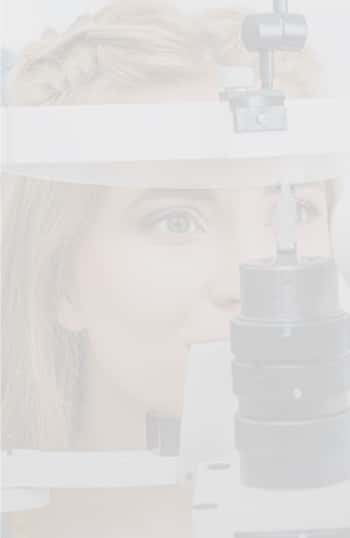
Sitting comfortably, you will rest your chin and forehead against the camera. We will then prepare and take the picture, with results available just a few moments after taking the picture.
Retinal imaging can show eye doctors a wide range of issues. These images can help with the diagnosis of the following problems:
- Retinal detachment
- Holes or tears in the retina
- Macular degeneration
- Eye damage arising from high blood pressure, arteriosclerosis or diabetes
- Glaucoma
- Age-Related Macular Degeneration
- Retinal tears and detachments
- Optic strokes
- And many others
We also see signs of non-eye diseases, such as the development of diabetes or high blood pressure.


